all articles
Take
Action

Artificial Intelligence
May Be the Key to Beating Cancer

By Joe Van Brussel
Medically reviewed by Teresa Hagan Thomas PHD, BA, RN
Cancer is a uniquely challenging adversary. As cancer cells multiply and spread, they do so quietly — patients often don’t feel sick in early stages, and doctors don’t see anything problematic on scans or tests.

Then, once symptoms do start to appear and tumors become large enough to be detected, it’s often late in the process, and intervention is more difficult.

It’s not that the patient or the doctors or the treatments are at fault — it’s that the best time to fight cancer is usually before it causes any symptoms.

Scientists have even discovered recently that cancer’s deception goes further. Thanks to advanced technologies using genetic data and machine learning, researchers discovered that certain cancerous tumors find places in the body where our immune system is particularly vulnerable. Cancer not only evades detection, but it also seeks out our weak spots.

But humanity is fighting back. While cancer may be deceptive, our best doctors, scientists, and technologists are getting better at finding it sooner. And the earlier we find it, the better we become at defeating it.

“We can now see things we’ve never been able to see before,” says Jenny Yu, MD, FACS, senior manager of Medical Integrity at Healthline

The recent improvements in early detection are due in large part to advances in artificial intelligence (AI) and machine learning. While popular conceptions of these technologies bring to mind self-driving cars or autonomous robots, medical professionals see them more as powerful partners or targeted tools that can be deployed when needed.

“AI is a great opportunity to do what we do better,” says radiologist Elliot Fishman, MD, who serves as co-principal investigator of the Felix Project for Early Detection of Pancreatic Cancer. “We’re working hard, but we need help, and if you want to improve things substantially, you need to put everything together.” 

This powerful solution isn’t a hypothetical. It’s not wishful thinking or another research paper — it’s been proven to work.... We could be looking at a sea change in the fight against cancer.

Artificial intelligence and similar technologies, which process a large amount of information very quickly and categorize it in a way that’s useful for physicians, are particularly suited to “put everything together.” 

Algorithms can provide a huge boost to care, both from a visual perspective (analyzing CT scans or other images quickly and efficiently) and from a data perspective (cross-referencing multiple data sets and categories and looking for similarities or patterns). 

This powerful solution isn’t a hypothetical. It’s not wishful thinking or another research paper — it’s been proven to work. 

Fishman and institutions like Northwestern Medicine have witnessed the accuracy and effectiveness of machine-assisted detection and are now implementing it in the real world.

Should clinical trials and prototypes mirror initial successes, we could very well be looking at a sea change in the fight against cancer.

Better Than the Human Eye

When speaking with Mozziyar Etemadi, MD, PhD, one wouldn’t guess at his extensive academic background. He speaks without relying on jargon or overly technical terminology.

Yet his résumé is impressive: He has a master’s of science in electrical engineering from Stanford, a doctorate in bioengineering from UC San Francisco and UC Berkeley, and a medical degree from UC San Francisco. He’s now assistant professor of anesthesiology and engineering at Northwestern.

His background in a multitude of fields spanning engineering, medicine, and technology puts him in a unique position to take advantage of these powerful new tools. Also, he points out, his medical specialty, anesthesiology, is a discipline that collaborates with virtually every team in the hospital.

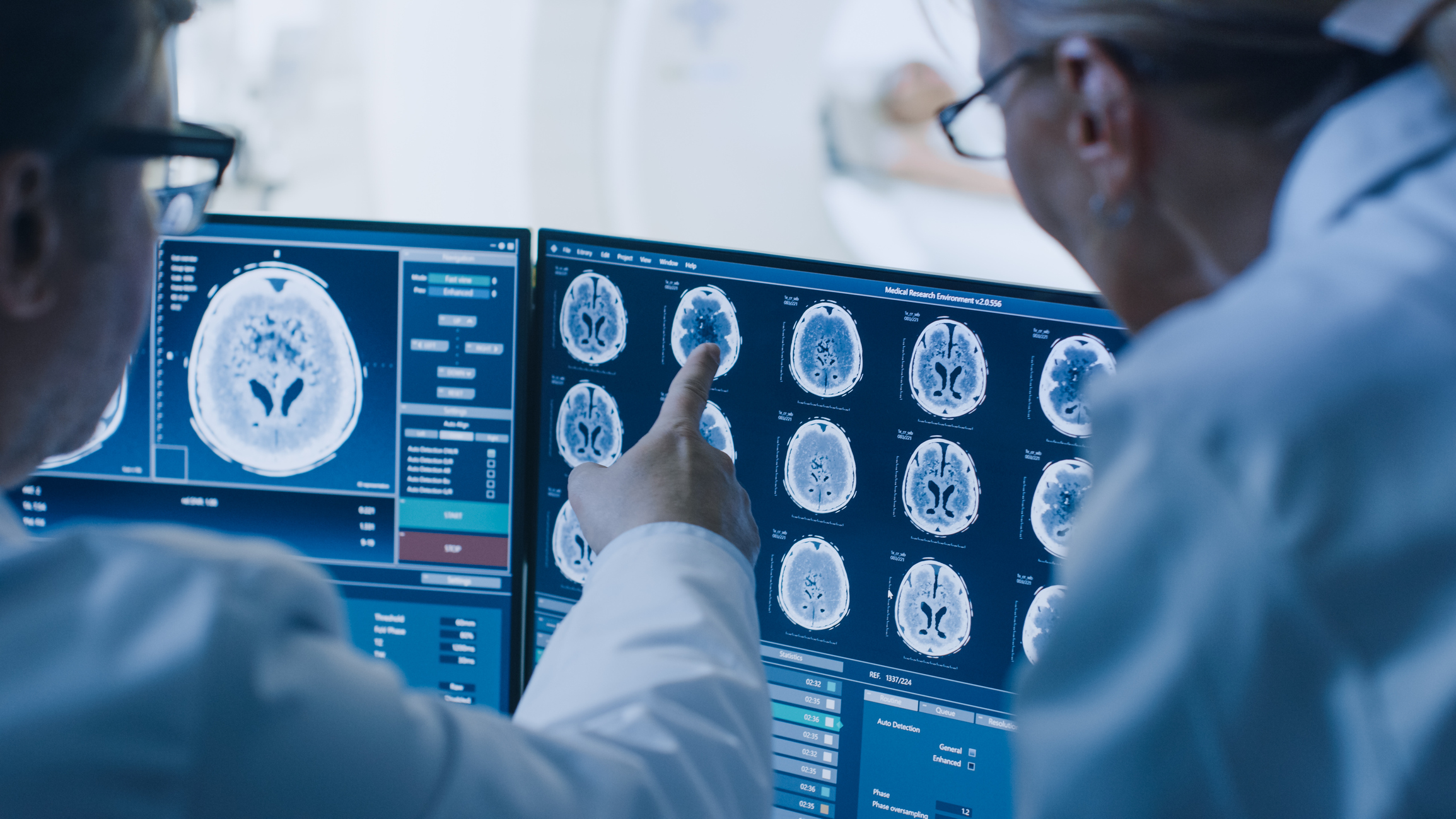
Etemadi and his colleagues fed their AI-based systems tens of thousands mammograms and CT scans until the computers were able to identify issues, on their own, in seconds.... with similar or better accuracy than the human eye.

“We work with surgeons, we work in the ICUs, we help deliver babies,” Etemadi says. “It’s a well-rounded foray, so when we’re developing these data tools, we have direct access to a lot of end users.”

So it perhaps wasn’t a surprise when the joint initiative he was working on for the last several years developed AI algorithms that use AI to read visual scans better than the human eye can.  That system is now being tested in the real world, with the possibility of a wider roll-out in the near future.

Working with Google and the U.K. National Health Service, Etemadi and his Northwestern Medicine colleagues fed their AI-based systems tens of thousands mammograms and CT scans until the computers were able to identify issues, on their own, in seconds. 

What’s more, the algorithms are able to identify problematic parts of an image with similar or better accuracy than the human eye. 

The team celebrated this massive milestone success, but they didn’t dwell on it. Before long, they were drawing up plans for a clinical study to test their systems in the real world. 

That trial is now running at Northwestern and will enable the algorithms to prove their merit not just retroactively when the outcomes are already known, but on a predictive basis as well. 

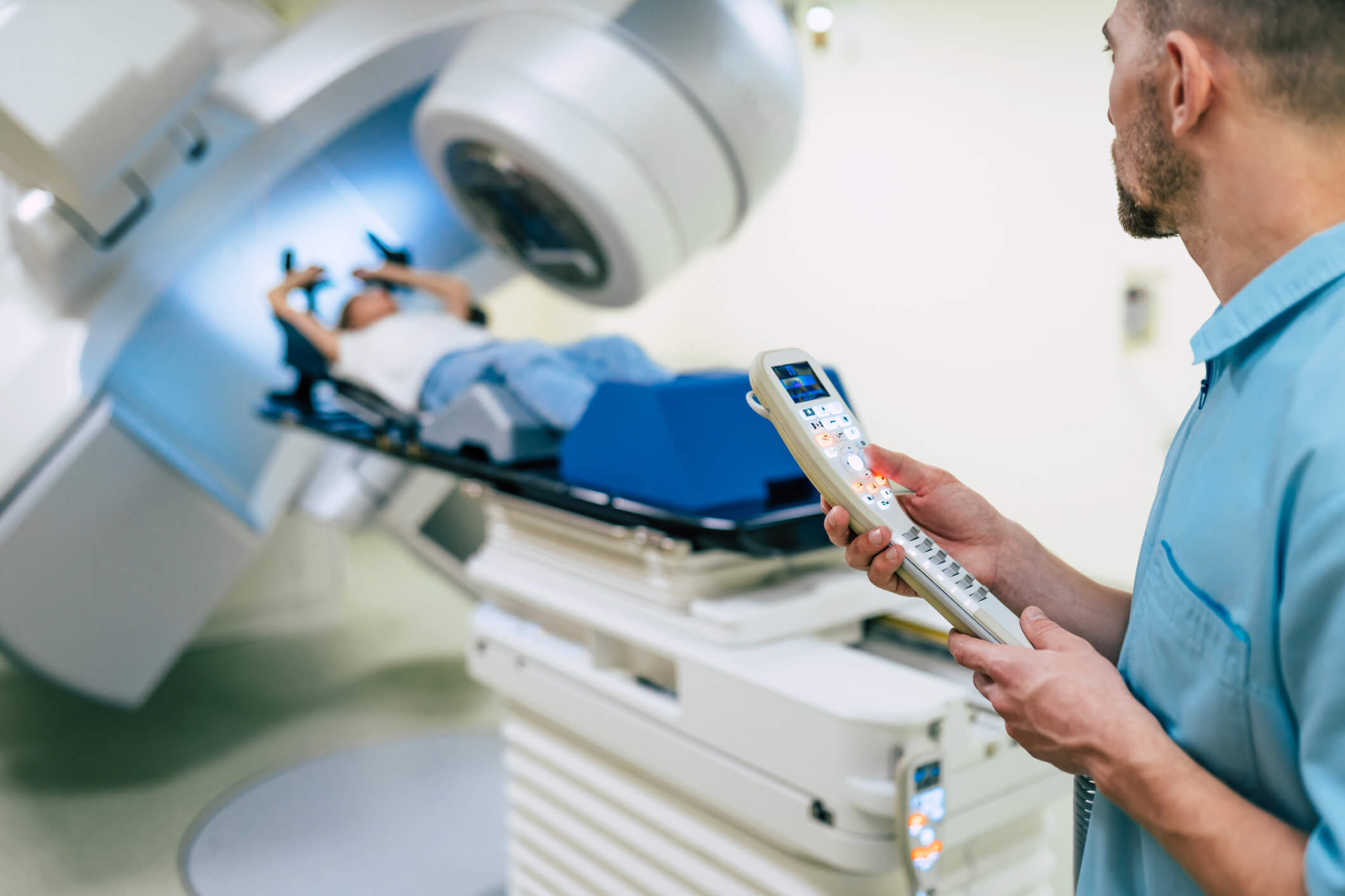
The study will focus on breast cancer and will completely transform the current process for mammograms and follow-ups. 

In the current system, patients have a mammogram done, then typically wait up to 30 days for it to be read by a specialist. If there are potential problems, the patient is contacted and they schedule a follow-up diagnostic mammogram, which “is a more intense mammogram essentially,” Etemadi says. 

Then, depending on the availability of radiologists and hospital resources, the patient waits again until physicians can review the diagnostic scan and settle on a diagnosis.  

But with the new algorithm, mammograms can be read by the computer within minutes. If a scan is flagged by the algorithm as high risk or problematic, the oncologist or radiologist can review it immediately and determine whether a diagnostic mammogram is necessary, before the patient has even gotten dressed. 

With improved speed and efficiency, patients will experience less wait time and anxiety. Some research suggests that AI-based scan reading could help relieve some “scan-xiety” patients have when waiting for scan results.

The “re-ordering of the queue,” as Etemadi describes it, allows radiologists to spend more of their time on suspicious scans and drastically speeds up the process for patients when it comes to the identification of tumors or abnormalities. 

The clinical study will also be key to understanding how the AI tools will be integrated and adopted by physicians, a task that’s just as critical as the effectiveness of the tools themselves.

“It’s really important to take time and make sure that the interaction between the human and the tool is as good as it possibly can be,” Etemadi says. 

With improved speed and efficiency, patients will experience less wait time and anxiety. Some research suggests that AI-based scan reading could help relieve some “scan-xiety” patients have when waiting for scan results.

With a smart tool like AI, radiologists will be able to focus more on critical scans, and their workload will be handled more efficiently.


That’s especially important given how high in demand cancer screening is today. And ultimately, patient outcomes may improve — more tumors will be caught, and they’ll be caught earlier.

Finding Hope

with One of the

Deadliest Cancers

Pancreatic cancer has a 5-year survival rate of about 10 percent, largely due to the advanced stage when it’s usually discovered.

Around 40,000 to 50,000 people die each year in the United States from pancreatic cancer.

“Some powerful treatments have come along, but it’s  not been as impactful as one would hope because it’s so late,” Fishman says. “The good news is that now you can detect early-grade disease and improve the survival rate.”

“When you can pick the cancer up early, you can change the survival curve,” Fishman explains. “Our focus is trying to bend the curve.”

Fishman and his colleagues at Johns Hopkins have also had success with AI-based algorithms. Their Felix Project is focused on pancreatic cancer. Pancreatic cancer has a 5-year survival rate of about 10 percent, largely due to the advanced stage when it’s usually discovered.

“Some powerful treatments have come along, but it’s not been as impactful as one would hope because it’s so late,” Fishman says. “The good news is that now you can detect early-grade disease and improve the survival rate.”

Fishman and the Felix Project team recently developed a working prototype that identifies abnormalities in the pancreas earlier than has ever been possible. The system uses AI to learn to spot tumors in CT scans before they’d normally be recognizable. 

At the beginning of its development, a team of radiologists and oncologists had to manually go through thousands of scans to label and identify aspects of a CT so the computer could learn. 

The democratization of the technology is key to the Felix Project’s mission and will help ensure that powerful detection tools aren’t limited to elite healthcare institutions or reserved only for those with means.

The prototype hasn’t yet seen enough cases for full approval, but “every case we’ve done so far, we’ve been accurate on,” Fishman says. And it’s easy enough to use that a radiologist who isn’t extensively trained in computer science likely won’t have issues.

Now the team is focused on expanding the number of cases and fine-tuning the prototype such that it can be used broadly. 

The democratization of the technology is key to the Felix Project’s mission and will help ensure that powerful detection tools aren’t limited to elite healthcare institutions or reserved only for those with means.

“We want to make certain that no matter who’s using it, it works well,” Fishman says. 

Around 40,000 to 50,000 people die each year in the United States from pancreatic cancer. Scientists believe causes may include the aging population, smoking, alcohol intake, and increases in obesity and diabetes, along with genetic risk factors.

When you can pick the cancer up early, you can change the survival curve. Our focus is trying to bend the curve.
Elliot Fishman, MD, FACS
Co-principal investigator, the Felix Project for Early Detection of Pancreatic Cancer

Given the difficult location of the pancreas — it sits between your stomach and your spine, and it’s surrounded by your gallbladder, liver, and spleen — detection is particularly difficult, even with high quality images. 

Technology like the prototype developed by the Felix Project could change the prognosis for countless patients across the country and globe. It may allow oncologists and surgeons to operate before the cancer has spread too far and save the lives of thousands of people.

“When you can pick the cancer up early, you can change the survival curve,” Fishman explains. “Our focus is trying to bend the curve.”

Looking

Ahead

While the advances in artificial intelligence and machine learning have been essential to the remarkable progress in early detection recently, doctors and technologists agree that it’s the collaboration between physicians and these new tools that’s most important. A tool is only as effective as its user and its application. 

Just as autopilot didn't replace the pilot, ...it's really a marriage of human and machine that allows these leaps in safety to occur.
Mozziyar Etemadi, MD, PhD

Even now, as clinical trials and prototypes show progress, there’s little talk of a world in which computers provide an actual diagnosis on their own. They can, however, vastly improve accuracy and safety.

Just as autopilot didn't replace the pilot, “it's simply an effective tool for helping the pilot get the passengers where they need to go as safely and smoothly as possible,” explains Etemadi. “It's really a marriage of human and machine that allows these leaps in safety to occur.”

And clinicians and technologists are already thinking about the future. While algorithms like the ones used by Etemadi and Fishman are now trained to look for cancer in a specific organ or region, new deep learning techniques will allow systems to find patterns and problems regardless of the type of cancer or part of the body. 

Teams like the Felix Project are also looking at ways of deploying AI and machine learning to use multiple sets of data, not just visual scans.

“We've been working on using a combination of imaging data as well as lab data and other clinical data, to be able to look and predict where to find pancreatic lesions,” Fishman says. 

Such investigations could move the field forward significantly when it comes to identifying higher risk patients.

All these advances point to a future when providers will be able to face cancer on a more favorable battlefield. “All warfare is based on deception,” the Chinese general and philosopher Sun Tzu once said. 

Thanks to these pioneers, cancer’s deception could soon be thwarted.

Keep Exploring

Stay on top of the latest developments in health and medicine with this daily dose of news stories.

Thank you! Your submission has been received!
Oops! Something went wrong while submitting the form.
share